Calvarial Hyperostosis in Lions
The Lion (Panthera leo), was once abundant over most of the African continent, vast areas of Asia and even Europe. Having two subspecies; the African and Asiatic lions, are facing grave danger of extinction due to habitat loss, illegal poaching and persecution. The current geographical range and lion population have frightfully decreased and is now considered at a vulnerable - VU (African subsp.) and endangered - EN (Asiatic subsp.) conservation status by the International Union for Conservation of Nature (IUCN) [1]. Several hundreds of lions are reported to be domiciled in captivity, with their role in the future conservation of their species yet to be determined.
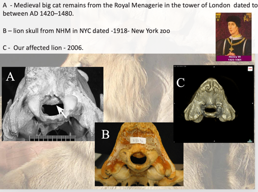
Lions have been kept in zoological gardens worldwide for decades and are considered the highlight in many parks. They seem to adjust to captivity well and usually are easy to reproduce. Therefore, lions are not at the center of attention when zoological gardens are asked to take part in conservation programs. Nevertheless, it is well documented that stillbirth, cub’s and young adult’s death due to neurological dysfunction are frequently seen in many zoological gardens around the world. Most of the reports are related to Calvarial Hyperostosis which results in reduced volume of the caudal fossa that causes compression of the cerebellum and spinal cord leading to severe ataxia, discomfort and finally death. Similar bone pathology was found in medieval big cat remains dated 1420-1480 and is still found in captive lions these days.
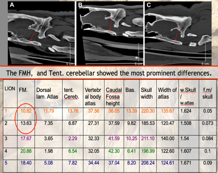
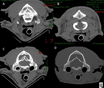
Our group has been looking at ways to achieve diagnosis, treatment and prevent the occurrence of Calvarial hyperostosis in captive lions. Through the years we were approached by zoo and private veterinarians from different parts of the world, looking for advice regarding the diagnosis and treatment of neurologically affected lions. Although vitamin A deficiency is believed to be the underlying cause, our findings could not rule out genetic and other environmental factors that may predispose certain lions to develop the abnormal bone growth, while others in the group remain healthy. Although the disease has been so far documented in captive lions primarily, further understanding of its precise pathophysiology is needed before it is guaranteed to not be a possible risk for the health of the entire lion population.We have documented the bone pathology characterizing calvarial hyperostosis using CT of healthy (Figure 2, B & D) and affected lions (Figure 2, A & C) and published guidelines for the diagnosis of this condition in lions (Figure 3).
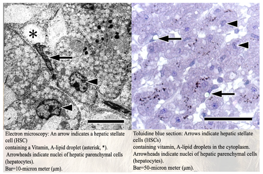
We further developed a safe way to measure liver retinoic acid concentration to monitor the lion’s status in regard to this important vitamin using ultrasound guided tru-cut needle biopsy as shown in the picture (Figure 4). Supplementing lions with the vitamin has shown to ameliorate clinical signs if given early in the course of the disease. We also demonstrated using electron microscope (EM) that hepatic stellate cells containing vitamin A lipid droplets are abundant in captive lions with very low serum vitamin A concentration.
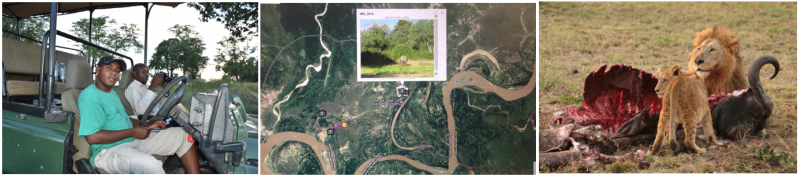
It is well accepted that vitamin A is needed through early stages of fetal development to allow normal morphology and development of the skull, more specifically the caudal fossa. It is therefore expected that pregnant lioness must have sufficient amount of vitamin A to provide the need of two and sometimes four cubs, possibly more than the rest of the lion pride. We are currently looking for differences in eating behavior of members of the pride in the wild to help estimate the amount of vitamins (A and others) consumed by each of them. This project is run by local Safari guides who observe and document the behavior during their daily work. Billy Nkhoma is heading the project in Zambia.
References:
-
Shamir, H.M., Horowitz, I.H., Yakobson, B., Ofri, R., (1998) Arnold-Chiari malformation in a captive African lion cub. J Wildl Dis.; 34:661-6. abstract
-
Shamir, H.M., Shilo, Y., Fridman, A., Chai, O., Reifen, R., Miara, L. (2008). Sub-occipital craniectomy in a lion (Panthera leo) with occipital bone malformation and hypovitaminosis A. J Zoo Wildl Med, 39:455-9. abstract
-
Gross-Tsubery, R., Chai, O., Shilo, Y., Miara, L., Horowitz, I.H., Shmueli, A., Aizenberg, I., Hoffman,C., Reifen, R., Shamir, H.M. (2010). Computed tomographic analysis of calvarial hyperostosis in captive lions. Vet Radiol Ultrasound. 51:34-8. abstract
-
Shamir, H.M., Rubin, G., Aizenberg, Z., Berkovitz, Z,. Reifen, R., Horowitz, I., Bdolah-Abram, T., and Aroch, I., (2012). Needle biopsy for hepatic vitamin A levels in lions (Panthera Leo). J. Zoo Wildl Med. abstract
-
Saragusty, J., Nadler, R., Shavit-Meyrav, A ., Yamaguchi,N., Bdolah-Abram,., Gibeon, L., Shamir, H.M. (2014). Comparative Skull Analysis Suggests Species-Specific Captivity-Related Malformation in Lions (Panthera leo). PLoS One, 2014 Apr 9(4):e 94527. abstract
-
Bauer, H, Nowell K. & Packer C. Panthera leo, In: IUCN Red List of Threatened Species, Version 2013. abstract
-
Clubb R, Mason G (2003) Animal welfare: Captivity effects on wide-ranging carnivores. Nature 425: 473-474. doi:10.1038/425473a. abstract
-
O’Sullivan BM, Mayo FD, Hartley WJ (1977) Neurologic lesions in young captive lions associated with vitamin A deficiency. Aust Vet J 53: 187–189. abstract
-
O’Regan HJ, Turner A, Sabin R (2006) Medieval big cat remains from the Royal Menagerie at the Tower of London. Int J Osteoarchaeol 16: 385–394.doi: 10.1002/oa.835. abstract
-
Chandra AMS, Papendick RE, Schumacher J, Homer BL, Wollenman P (1999) Cerebellar herniation in captive lions (Panthera leo). J Vet Diagn Invest 11: 465–468. abstract
-
Bartsch RC, Imes GD Jr, Smit JPJ (1975) Vitamin A deficiency in captive African lion cubs (Panthera leo) (Linnaeus, 1758). Onderstepoort J Vet Res 42: 43–54. abstract
-
Barnett R, Yamaguchi N, Barnes I, Cooper A (2006) Lost populations and preserving genetic diversity in the lion Panthera leo: Implications for its ex situ conservation. Conserv Genet 7: 507–514. doi: 10.1007/s10592-005-9062-0. abstract
-
Burroughs RE, Roos CJ, Ebedes H (1988) Inco-ordination and paresis in a captive lion (Panthera leo). J S Afr Vet Assoc 59: 81–82. abstract
-
Zile MH (2001) Function of vitamin A in vertebrate embryonic development.J Nutr 131: 705–708. abstract
-
McCain S, Souza M, Ramsay E, Schumacher J, Hecht S, et al. (2008) Diagnosis and surgical treatment of a Chiari I-like malformation in an African lion (Panthera leo). J Zoo Wildl Med 39: 421–427. doi: 10.1638/2007–0157.1. abstract
-
Hartley, M. P., R. M. Kirberger, M. Haagenson, and L. Sweers. 2005. Diagnosis of suspected hypovitaminosis A using magnetic resonance imaging in African lions (Panthera leo). J. South Afr. Vet. Assoc. 76: 132–137. abstract