check
Micro CT - Scanco | Core Research Facilities

Micro CT - Scanco

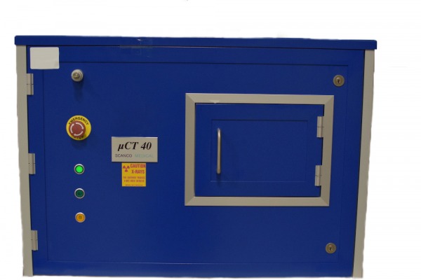
Computed tomography (CT) has both clinical and pre-clinical applications, using X-ray beams in order to record data regarding the anatomy and density of specimens or living tissue. Micro-CT (μCT) technology enables to obtain information regarding the microstructure of organisms.

Our Scanco μCT 40 can scan images of ex-vivo specimens with a resolution of 8 μm voxel sizes.

The procedure occurs in 3 steps:

  1. Raw image acquisition by the Scanco μCT 40 machine.
  2. Rapid image reconstruction in the axial plane by an HP Z840 Workstation with a dedicated Graphics Processing Unit (GPU).
  3. User performed analysis done on an HPE Integrity rx2800 i4 tower.


Prior to usage, a mandatory instruction is required please contact us.


Link on manufacture’s website


Specifications

µCT 40

Type

Desktop cone-beam microCT
Fully shielded
No external shielding required

X-ray source

Sealed
Air-cooled
Maintenance-free
30 - 70 kVp

Detector

2048 x 256 elements, 24 µm pitch 

Resolution

‹ 8 µm 10% MTF
3 - 72 µm nominal isotropic (pixel size)

Image matrix

512 x 512 to 4096 x 4096 pix

Max. scan size

36.9 x 80 mm (ØxL)

Max. specimen size

36.9 x 80 mm (ØxL)

Electrical

100 - 230 V / 50 - 60 Hz
345 W


MicroCT

line